Compact Bone Diagram Unlabeled : Art Labeling Activity Structure Of Compact Bone Diagram Quizlet - It can be remodeled all throughout life identify the circular vessels in the middle of bone running circumferentially around the vessels.
byAdmin-
0
Compact Bone Diagram Unlabeled : Art Labeling Activity Structure Of Compact Bone Diagram Quizlet - It can be remodeled all throughout life identify the circular vessels in the middle of bone running circumferentially around the vessels.. Skeletal system blank diagram diagram of anatomy. The long bones of the body contain many distinct regions due to the way in which they develop. The outer part of a long bone is made of compact bone. Compact bone is made of a matrix of hard mineral salts reinforced with tough collagen fibers. Start studying anatomy bone diagram long bone.
The outer part of a long bone is made of compact bone. Human anatomy physiologyil biol 1611l. Human gross anatomy study | humandiagram.info. Save the image and use it to replace the unlabeled one into your blog which major regions and structures of an osteon in a histological specimen of compact bone (or diagram or model of one) can you identify? Its unlabeled, so that your practce better.
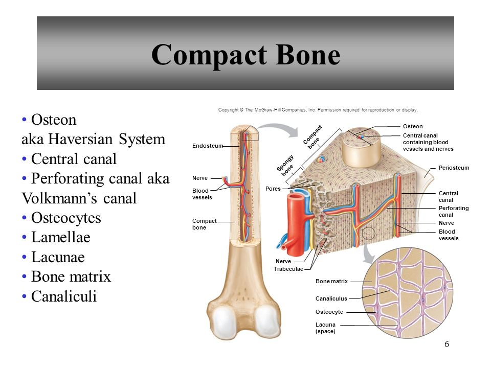
Chapter 7 Skeletal System Ppt Download from slideplayer.com Human anatomy physiologyil biol 1611l. It can be remodeled all throughout life identify the circular vessels in the middle of bone running circumferentially around the vessels. The compact bone is the main structure in the body for support, protection, and movement. This shows the architecture of compact bone which is designed to nourish and regulate osteocytes and bone matrix. Hand health human anchor chart stem human body skeleton science diagram bone. The outer part of a long bone is made of compact bone. Unlabeled diagram showing the carpal bones (download free pdf below!) now you've seen the carpal bones labeled and unlabeled, it's time to move on to our interactive carpal bones quizzes. What is the difference between compact and spongy bone?
Terms in this set (23).
The outer part of a long bone is made of compact bone. Muscles of the body identification7 games. It can be remodeled all throughout life identify the circular vessels in the middle of bone running circumferentially around the vessels. Cervical vertebrae blank bone diagram skeleton quiz diagrams. Bone long anatomy epiphysis marrow structure diaphysis epiphyseal human leg metaphysis osteoporosis trabecular vector yellow anatomical biology blood body care cartilage cavity compact diagram education educational femoral femur fibula health health care healthy illustration line medical. The skeletal system is a topic of the event anatomy for the 2020. What is the difference between compact and spongy bone? Quizzes on human skeletal system anatomy, bone anatomy, and bone markings. Save the image and use it to replace the unlabeled one into your blog which major regions and structures of an osteon in a histological specimen of compact bone (or diagram or model of one) can you identify? Carotid canal coronal suture ethmoid bone external occipital protuberance foramen lacerum foramen magnum foramen. Advanced skull labeling free worksheets google search skeleton co. The tibia (os tibia) and fibula (os fibula) are the bones. Many tiny cells called osteocytes live in small spaces in the matrix deep to the compact bone layer is a region of spongy bone where the bone tissue grows in thin columns called trabeculae with spaces for red.
The skeletal system is a topic of the event anatomy for the 2020. Start studying anatomy bone diagram long bone. Compact bone high resolution histology diagram. Its unlabeled, so that your practce better. Structure of long bones dra.
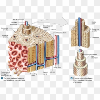
Compact Bone Microscopic Labeling Diagram Quizlet from o.quizlet.com Compact bone consists of outer and inner sheets of lamellar bone (not seen here) and haversian systems, shown here, that run parallel to the long axis of bones. Start studying fracture repair unlabeled. Learn vocabulary, terms and more with flashcards, games and other study tools. Part 3 microscopic structure of compact bone. Terms in this set (23). Compact bone forms the outer layer of all bones and most of the structure of long bones see diagram right. Begin by identifying the concentric rings of lamellar bone that surround a haversian canal. Practice quiz & test prep for students and teachers.
Skeletal system blank diagram diagram of anatomy.
Terms in this set (23). Begin by identifying the concentric rings of lamellar bone that surround a haversian canal. Compact bone is made of a matrix of hard mineral salts reinforced with tough collagen fibers. Start studying fracture repair unlabeled. Cervical vertebrae blank bone diagram skeleton quiz diagrams. Microscopic bone anatomy human body diagram. Cancellous bones, compact bone, cortical bone, diaphyses, haversian canal, lamella, marrow cavity, osseous tissue, osteons. The compact bone is the main structure in the body for support, protection, and movement. The skeletal system is a topic of the event anatomy for the 2020. Learn vocabulary, terms and more with flashcards, games and other study tools. Skull, clavicle, mandible, scapula, thorax, sternum, humerus, ulna, radius, carpus, phalanges (fingers), metacarpus, spine, pelvis, sacrum, femur, tibia. Long, short, flat, irregular and sesamoid. Human gross anatomy study | humandiagram.info.
There is a printable worksheet available for download here so you can take the quiz with nervous system cellular diagrams4 games. This shows the architecture of compact bone which is designed to nourish and regulate osteocytes and bone matrix. The outer walls of the diaphysis cortex cortical bone are composed of dense and hard compact bone a form of osseous tissue. Compact bone is the denser, stronger of the two types of osseous tissue (figure 6.3.6). The bones mentioned in each human skeleton chart are:
Bone Png Transparent For Free Download Page 2 Pngfind from spng.pngfind.com Sclerostin inhibits bone formation mostly by antagonizing lrp5/6, thus inhibiting wnt signaling. Compact bone high resolution histology diagram. Learn vocabulary, terms and more with flashcards, games and other study tools. Compact bone labeled diagram vmglobal co. The compact bone is the main structure in the body for support, protection, and movement. They run along the length. Due to the strong nature of compact bone, compared to spongy bone, it is the. Terms in this set (23).
Bones diagram human bones diagram human skeleton diagram human.
Due to the strong nature of compact bone, compared to spongy bone, it is the. Unlabeled diagram showing the carpal bones (download free pdf below!) now you've seen the carpal bones labeled and unlabeled, it's time to move on to our interactive carpal bones quizzes. There is a printable worksheet available for download here so you can take the quiz with nervous system cellular diagrams4 games. What are diplo , its function, and location? The structure of a long bone allows for the best visualization of all of the parts of a bone figure 1. Start studying anatomy bone diagram long bone. Learn vocabulary, terms and more with flashcards, games and other study tools. Quizzes on human skeletal system anatomy, bone anatomy, and bone markings. Structure of long bones dra. Gallery long bone diagram unlabeled anatomy and physiology. Begin by identifying the concentric rings of lamellar bone that surround a haversian canal. Compact bone consists of outer and inner sheets of lamellar bone (not seen here) and haversian systems, shown here, that run parallel to the long axis of bones. A typical long bone showing gross anatomical features.
The long bones of the body contain many distinct regions due to the way in which they develop compact bone diagram. The skeletal system is a topic of the event anatomy for the 2020.